literature review · co-authored with Jasper Fiscus

Applications of hydrogels in microfluidic devices

A review of how hydrogels are fabricated and used across microfluidic cell culture, drug delivery, and biosensing — and where the field's biggest limitations still sit.

Hydrogels & history

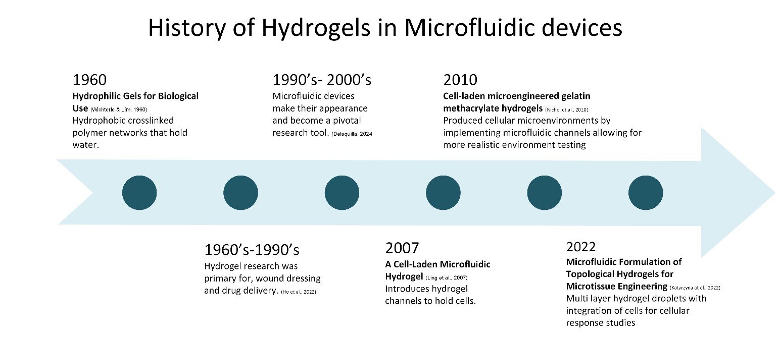
Hydrogels are water-rich, cross-linked polymer networks — made from natural materials like collagen, synthetic polymers like PEG, or blends like GelMA — prized for biocompatibility and tunable mechanical properties that let them mimic native extracellular matrix. Their use in microfluidics has grown fast: published research on the topic rose from around 4,000 papers in 2017 to over 6,500 in 2021, and keeps climbing.

The field traces back to 1960, when Wichterle and Lim introduced the first hydrogel for biological use. Early research through the 1990s focused on wound dressings and drug delivery; once microfluidic devices emerged as a research tool in the late '90s and 2000s, hydrogels and microfluidics began to merge, culminating in cell-laden microfluidic hydrogels by 2007 and today's organ-on-a-chip platforms.

Timeline infographic showing the history of hydrogels in microfluidic devices from 1960 hydrophilic gels through 2022 multi-layer hydrogel droplets.
History of hydrogels in microfluidic devices, 1960–2022

Fabrication methods

Four fabrication approaches dominate the field, each suited to different device geometries and throughput needs.

Molds — PDMS via photolithography, the most common method
Droplets — oil slipstream around an outlet tube produces hydrogel spheres
Fibrous spinning — glass tubes assembled into multi-layer strands
3D bioprinting — custom-built printers feeding multiple materials at once

Applications

Current research clusters into four categories: 2D and 3D flow cell culture, drug delivery, and sensors. A University of Wisconsin–Madison team, for example, used PEG-encapsulated hydrogels to model the tumor microenvironment in glioblastoma cells under controlled perfusion — capturing diffusion-limited drug transport that a flat Petri dish can't replicate.

Diagram of hydrogel loading for an angiogenesis coculture model and a simulated concentration contour of diffusive transport across hydrogel channels.
Loading hydrogels for an angiogenesis coculture model, and the resulting diffusive transport simulation (Lee et al., 2022)

On the drug delivery side, a University of British Columbia group built an implantable system using thermo-reactive hydrogel microvalves triggered wirelessly by radio frequency — releasing drugs on demand without any onboard power supply.

Diagram of an RF magnetic field activated microvalve drug delivery device and its working principle graph.
RF magnetic field-activated hydrogel microvalves for on-demand drug release (Rahimi et al., 2010)

Sensing applications range from pH-responsive photonic crystal beads to hydrogel-immobilized enzymes that detect glucose and galactose in real time — all leveraging the same core property: a hydrogel's structure changes measurably in response to its chemical environment.

Limitations & future work

The two biggest limitations we identified are fabrication difficulty and infrastructure cost. Molds don't allow fine spatial control; 3D bioprinting offers better resolution but suffers from scalability and reproducibility issues. On top of that, the cleanroom and cell-culture infrastructure needed to support this research is expensive to build and maintain.

cleanroom construction cost

$200–1,000/ft²

cell culture equipment cost

$25K–100K

We expect the most meaningful progress to come from more complex organoid- and multi-organ-on-a-chip devices, cheaper off-the-shelf 3D bioprinters, and further integration of hydrogels as responsive sensing matrices for wearable and point-of-care diagnostics.

key words

hydrogels microfluidics lab-on-a-chip organ-on-a-chip tissue engineering drug delivery biosensors literature review cell culture

Full review paper

Complete literature review with all figures, tables, and 52 references.

Download PDF